
Description
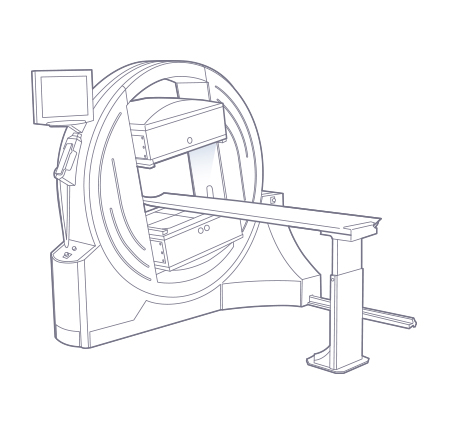
The nuclear imaging system is a versatile dual-detector single-photon emission computed tomography system for examinations of skeletal bones and internal organs. In the field of nuclear medicine, single-photon emission computed tomography is the most common and popular diagnostic method, which has proven to be useful due to its high quality, affordability, and cost-effectiveness. The method is the most relevant in oncology, cardiology, neurology, endocrinology, nephrology practice, etc.
The nuclear imaging system allows diagnosing diseases at early stages, at the molecular level, revealing both the primary focus and the extent of the process, assessing treatment efficiency, detecting recurrences. The nuclear imaging system is widely used and allows investigating various biological processes simultaneously during a single diagnostic procedure.
The nuclear imaging system has been developed taking into consideration the Russian nuclear medicine practice. Diagnostic procedures can be performed using the entire range of available radiopharmaceuticals. Their production does not require a cyclotron, which ensures their wide availability.
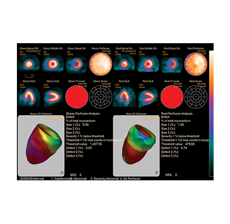
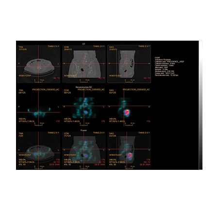
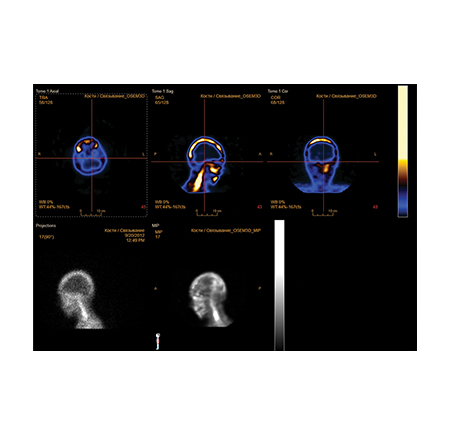
The nuclear imaging systems have the multilingual interfaces and are fitted out with specialized software to analyze the heart, brain, skeleton, liver, kidneys, esophagus, thyroid, parathyroid glands, and other organs. The availability of the combination function of images obtained using the SPECT manufactured by NIPK Electron Co. and computed tomography systems of any other manufacturer will make it possible for a radiologist to determine the localization and the extent of a pathologic process with the highest possible accuracy.
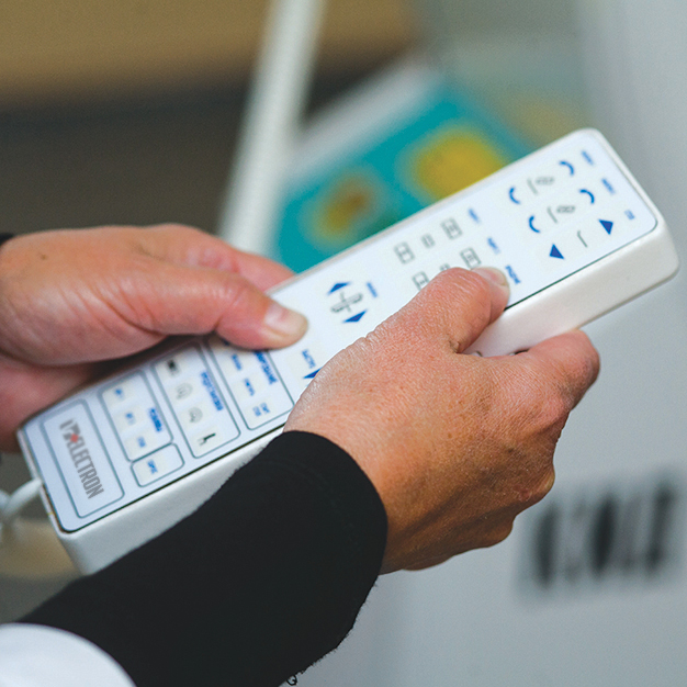
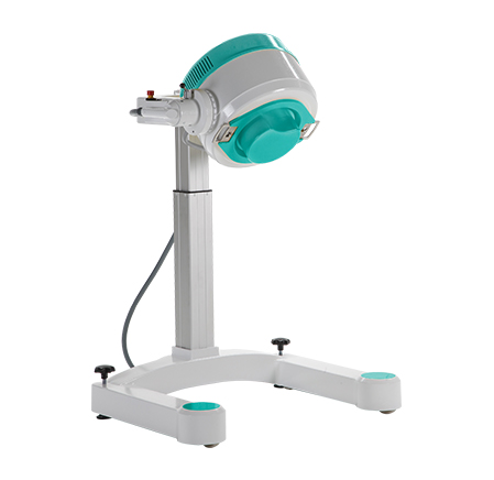
The nuclear imaging system is easy-to-use and user-friendly. The imaging process is fully automated; system initialization is performed by pressing one button. The stand design allows scanning the patient with height of more than 2 m what is of special importance in skeletal bones scintigraphy.
The nuclear imaging system has small overall dimensions, occupies only 6 sq. m, which makes it possible to install it in a room, in which equipment replacement is planned, as well as to establish an isotope diagnostics department even in case of free area deficiency.
Benefits
High diagnostic quality
-
Highly-sensitive digital detectors
- Examination result processing and analysis software harmonized with Russian practice
- Broad collimator nomenclature
Easiness-to-use and user-friendliness
-
Multilingual control station interface and software
- Complete imaging and calibration process automatization
- Fully automatic patient circuit construction system
- The use of the entire radionuclides nomenclature of Russian and foreign make
- Large table top height variation range
Safety
- Built-in system control and setup protocols as per standard NEMA NU 1 and GOST-R IEC 61675-2, GOST-R IEC 61675-3
- Collision prevention dual-circuit safety system
- Uninterruptible power supply allowing to complete the examination in case of power shutdown
Multifunctionality, small size
-
Examination of the patients in the supine, sitting, and upright position
- Specialized software to analyze the heart, brain, skeleton, liver, kidneys, esophagus, thyroid, parathyroid glands, etc.
- Examination types supported: planar (dynamic and static) examinations, SPECT (3D-reconstruction), ECG-synchronized cardiac imaging, whole body examination mode
- SPECT- and CT-image combination option
- Small-size (6 sq. m)
Reliability and durability
-
Simple mechanical components
- Low power consumption of the system
- Power line surge protection
Basic configuration *
Description
Automated workstations (AWS)
- Operator’s and radiologist’s automated workstations with a specialized software package for image analysis
- Additional radiologist’s automated workstations
- PACS server
Collimator set
- Low-energy general purpose collimators
- High-resolution low-energy collimators
- Medium-energy general purpose collimators
- High-energy collimator
Equipment of cardiological examinations
- Cardiac synchronization system
- Cardiovascular machine
Fitting out of a room and laboratory
- Uninterruptible power supply for gantry and automated workstations
- Climatic system
- Dose isotope calibrator
- Equipment for fitting out a laboratory for the work with radionuclides
- Patient view/position means
X-ray radiation protective equipment for patients and staff
- X-ray protective clothes for the medical staff
- Personal protection equipment for patients
- X-ray protective windows of different sizes
NIPK Electron Co. offers the following nuclear medicine systems types:
- Mobile planar gamma-camera with the 25 cm field of view
- Single detector SPECT system
- Dual detector SPECT system
- Three-detector SPECT system for the heart and brain diagnostics

*The “Basic configurations” section includes the information only on the most demanded configurations; the general list is significantly wider. Any configuration and technical parameters can be changed by the manufacturer’s initiative or at the customer’s request.
Do you have any questions?
Saint Petersburg: +7 (812) 325-02-02