
Description
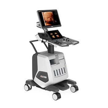
The many-year experience and competencies of NIPK Electron Co. in the field of design and production of medical imaging systems and high-technology equipment allowed designing a wide range of ultrasound systems of different classes: from medium to expert level.
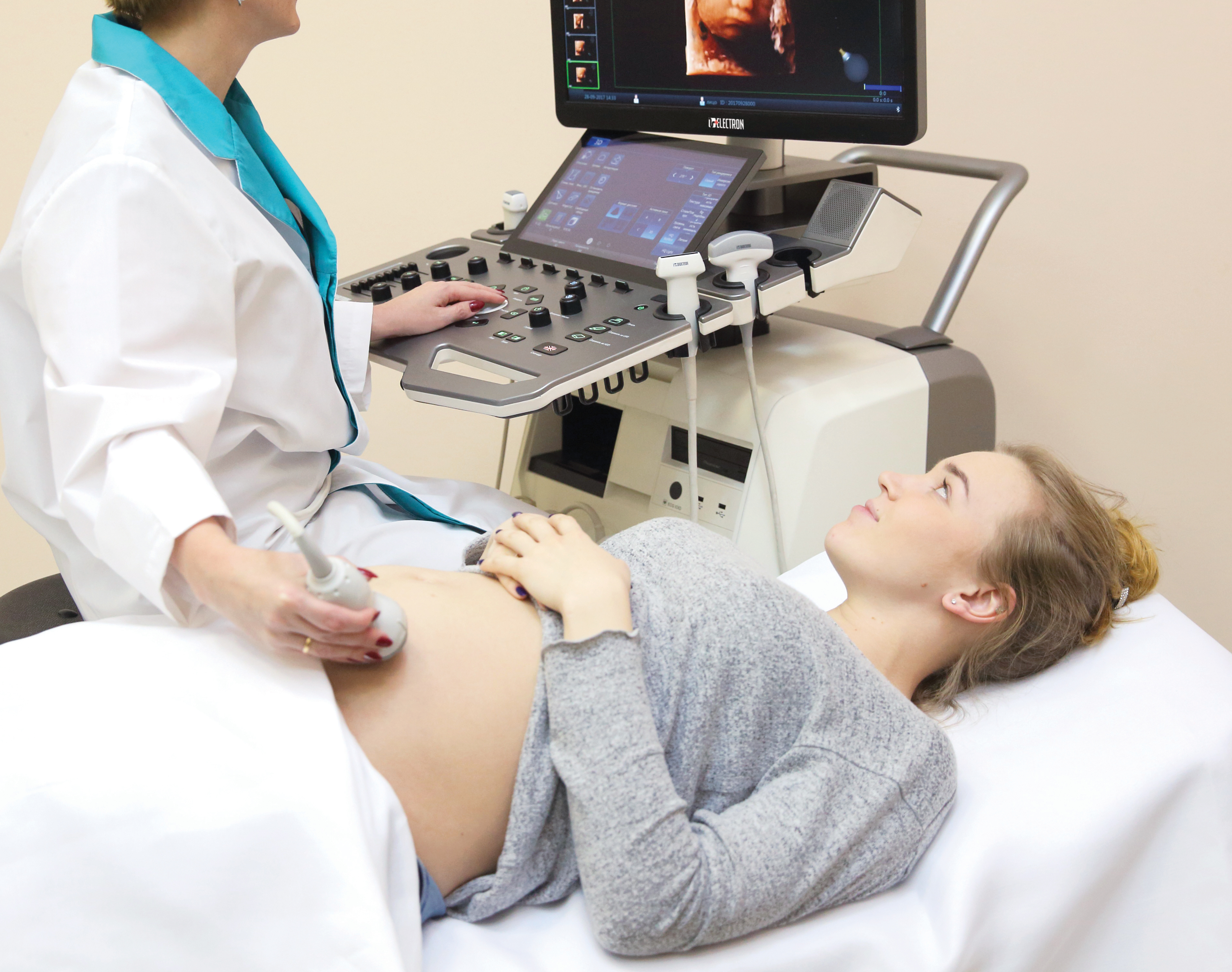
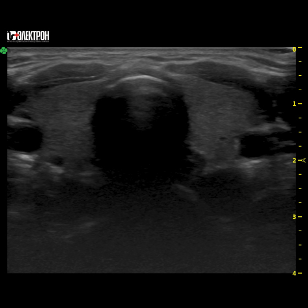
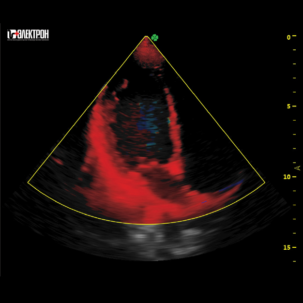
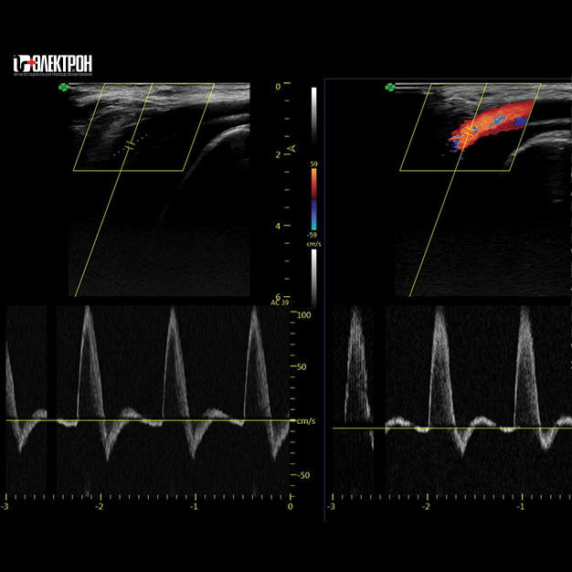
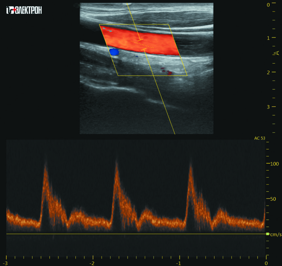
The diagnostic ultrasound systems manufactured by NIPK Electron Co. have a wide scope of application and are intended for abdominal, vascular, cardiology examinations; they are used in pediatrics, neonatology, obstetrics, and gynecology, for assessment of the musculoskeletal system, small and superficial organs, etc.
All equipment of the NIPK Electron are included in the unified register of Russian radioelectronic products (REP) (in accordance with the decree of the Government of the Russian Federation dated July 10, 2019 No. 878) and in the register of Russian industrial products (in accordance with the decree of the Government of the Russian Federation dated July 17, 2015 No. 719)
All equipment of the NIPK Electron are included in the unified register of Russian radioelectronic products (REP) (in accordance with the decree of the Government of the Russian Federation dated July 10, 2019 No. 878) and in the register of Russian industrial products (in accordance with the decree of the Government of the Russian Federation dated July 17, 2015 No. 719)
The scanners of the ultrasound systems range feature the ergonomic design and are fitted out with touch control panels. The systems of high and expert class allow regulating the operating arm and monitoring position for the specialist’s comfortable work. The ultrasound system can have probes with different frequency ranges including high-frequency linear probes of up to 18 MHz for certain examination types. The system can be configured both with standard transducers (piezo ceramic transducers) and monocrystalline transducers.
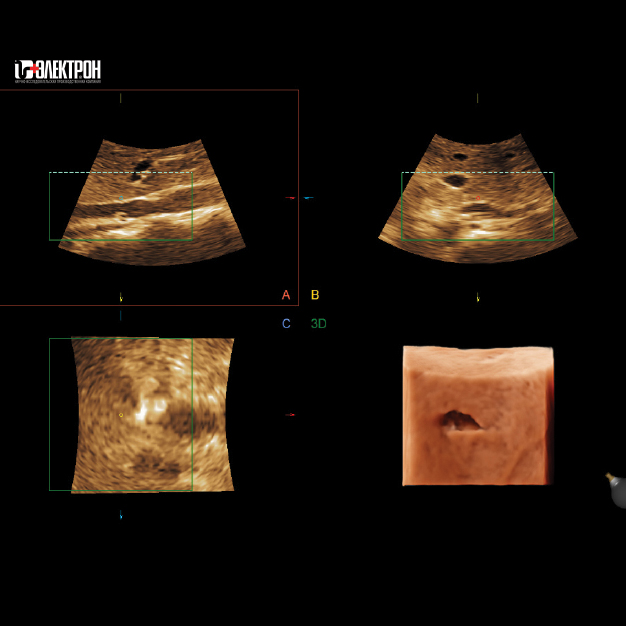
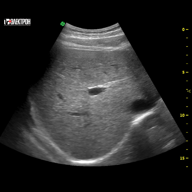
A unique post-processing system of the ultrasound system manufactured by NIPK Electron Co. allows processing data not subjecting it to additional demodulation, which makes it possible to maximize image clarity and contrast.
The software includes the full post-processing image improvement algorithm package aimed at granularity reduction, border enhancement, harmonic imaging, spatial and frequency compounding; the needle visualization improvement function is available for performing a puncture or a biopsy.
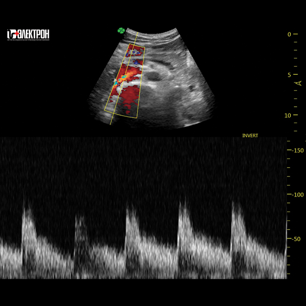
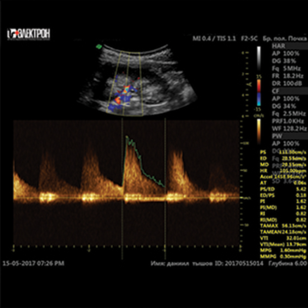
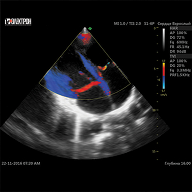
The ultrasound systems produced by NIPK Electron Co. support all data acquisition modes: B- and M-mode, color doppler, pulse wave doppler, power doppler and power-directed doppler, and tissue doppler. The data can be presented in duplex and triplex modes.
The software can also include echo-contrast agent packages, elastography, real-time image volume reconstruction, panoramic view. The images obtained using the ultrasound system and protocols can be saved on USB, DVD, shared via Wi-Fi and Bluetooth.
Any system can be configured with different programs and transducers for specific tasks — for cardiology, examinations of the musculoskeletal system, gynecologic examinations, performing interventional operations under ultrasound supervision.
Benefits
High diagnostic image quality
-
High image clarity and contrast
- Higher sensitivity of doppler imaging
- Real-time high-speed data processing rate
- State-of-the-art analysis of examination findings
- Automated program filters for image quality improvement
Easiness, simplicity, and user-friendliness
-
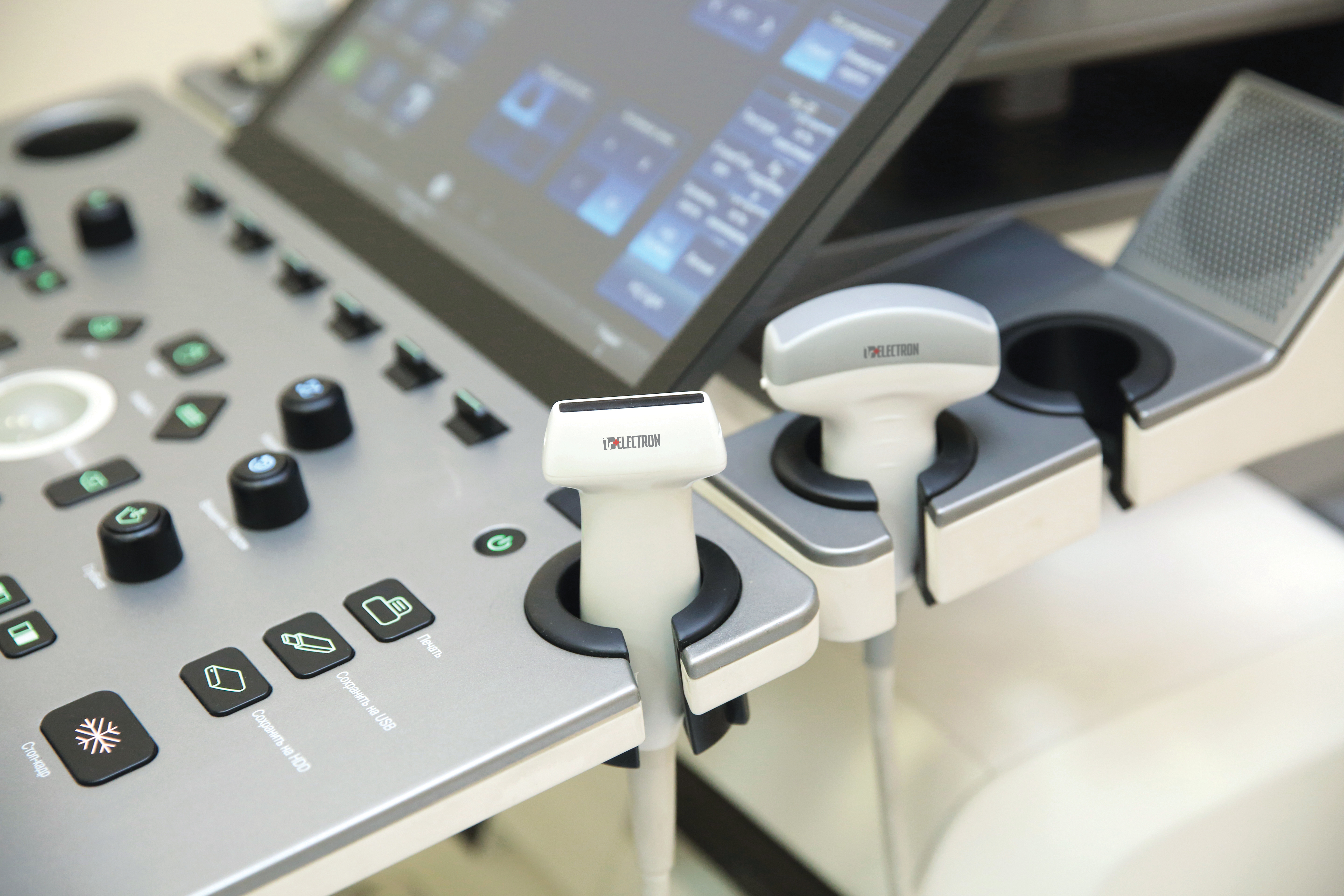
Touch control panel in all scanners
- Specialized rubberized holders for reliable transducer fixation
- User-friendly multilingual interface
- Possibility for examination result transfer to all types of external media (USB, DVD, external HDD)
- Data transfer via Bluetooth, LAN, Wi-Fi
- Sending images to the PACS
- DICOM image option
Mobility and multifunctionality
-
Adjustment of the arm position (height along and in horizontal plane)
- Monitor position adjustment
- From 4 to 5 transducer ports depending on the system class
- Gel heating function
- A wide transducer range including monocrystalline ones
Reliability and durability
- Elaborated, simple, and reliable system design
Basic configuration *
Description
Specialized software packages
- All types of dopplers (CFM, PWD, PDI, TD, CWD, TVI, TVM)
- M-Mode, Anatomical M-Mode, Color M-Mode
- Cardio package with ECG-gated image acquisition
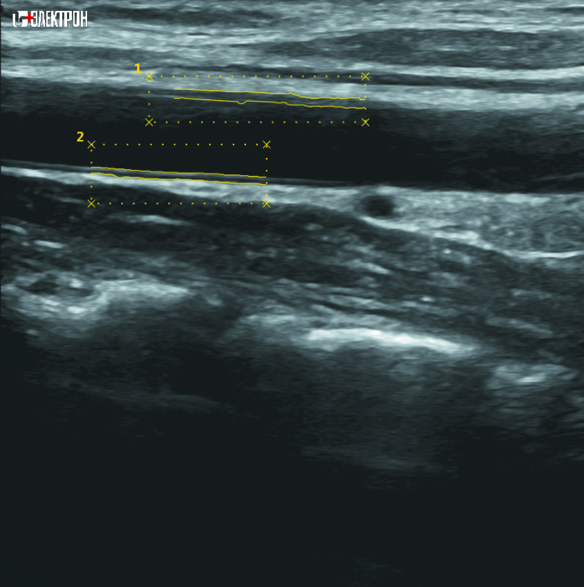
- Vascular package
- Obstetrics and gynecology package
- Pediatrics and neonatology package
- Kidney analysis package, urological package
- Panoramic view
- All modes of volume reconstruction (3D/4D)
- Contrast-enhanced imaging option
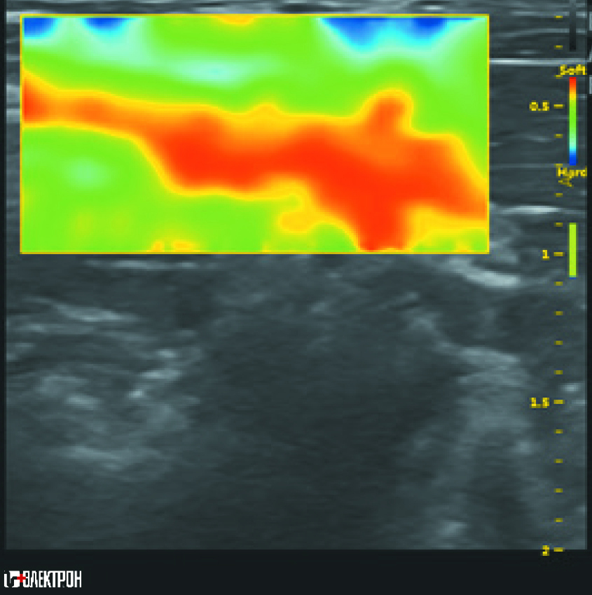
- Elastography
- Needle visualization improvement function for performing puncture or biopsy
- Implemented automatic measurements for various organs and systems
Additional equipment
- Image printer
- Uninterruptable power supply for the scanner
- Footboard set for scanner control
- PACS server
All latest technologies in different class systems*
- Five degrees of freedom tilt-swivel monitor bracket
- Gel heating
- Printer shelf
- Large area monitor
- Sensitive touch panel
- Up to 5 transducer ports
- Scanner control footboards
* The ultrasound systems manufactured by NIPK Electron Co. can be configured with different transducer types, specialized software packages, and periphery equipment. Technical parameters of the system can be changed by the manufacturer’s initiative or at the customer’s request.
Do you have any questions?
Saint Petersburg: +7 (812) 325-02-02
Expert opinions
All opinions