Description
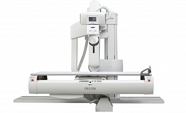
The versatile, fully automated system with a multi-functional fluoroscopy tilting table for multipurpose examinations ART is an essentially new tool of X-ray diagnostics with no analogues in the world, which opens up the maximum functional possibilities for users.
The multipurpose remote-controlled system is designed taking into consideration the needs of a wide range of radiologists based on the detailed analysis of the systems available on the world market and offers of the leading Russian experts in the field of diagnostic imaging. The system can be used instead of several other machines as it provides a wide range of diagnostic opportunities and allows performing various X-ray diagnostic examinations, in particular:
• Contrast-enhanced imaging of the gastrointestinal and genitourinary organs
• X-ray examinations of the thoracic organs and musculoskeletal system from the extremities up to the skull
• Interventional radiology procedures
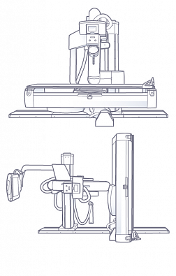
The unique system design provides for operation in any required mode: radiography, fluoroscopy, linear tomography, and with any necessary patient’s position (lying on the table or trolley including that in the lateral position, in the sitting or the upright position). The multipurpose X-ray tube stand moves around the patient making it possible to diagnose different injuries and diseases in any mode and at any angle from the patient’s head to feet This is of special significance in case of emergency examinations because it allows avoiding any additional injury and provides for rapid obtaining of important diagnostic data to ensure selecting the optimal treatment strategy.
The ART is equipped with a modern imaging system based on a large-format dynamic flat-panel detector that ensures the highest quality of obtained digital images.
A unique fluoroscopy processing system makes it possible to record a whole series of images or any fragment both remotely and directly at the X-ray table.
In addition to standard modes, the ART system features several progressive technologies in the field of image obtaining and processing such as:
• Tomosynthesis, which is a modern method of X-ray examination based on slice tomography image reconstruction of the while examined region from a sequential set of low dose angular views. This technology can be used successfully in diagnostics of pulmonary nodular lesions (including lung cancer), examinations of the musculoskeletal system, contrast-enhanced imaging of the GIT, urinary, etc. It allows not only to reveal the lesion but also ensures the determination of its accurate localization.
• Stitching: this method makes it possible to obtain and combine several images to create a panoramic view of the vertebral column or long bones during one examination in automatic mode what is very relevant when diagnosing musculoskeletal disorders, the degree of scoliosis as well as when planning surgical treatment.
The control system allows positioning the tube stand and performing the examinations completely remotely; in this situation, an X-ray technician can check the patient’s position using a video camera built-in in the collimator. The maximally flexible user interface of the control system allows a radiologist or a radiology technician to select the convenient settings, generate their own APR programs; set up the tomography parameters along with many other things.
If necessary, technical solutions used in the ART system allow implementing a remote connection in the online mode to diagnose and remove faults, and also set up the system according to with user demands.Benefits
Mobility and multifunctionality-
The widest range of system movement providing for the following:
• Imaging in lateral position both on a tilting table and a mobile X-ray table
• Imaging of the upper extremities (the humerus, the elbow joint, hands) directly to the detector
• Imaging of the lower extremities including weight-bearing foot radiography in lateral view and in dorsoplantar view
• Imaging in the oblique view on the table edge (imaging of cranial bones in oblique views, Mayer’s view, etc.)
• Chest radiography with the detector positioned either vertically or horizontally
• Examination of the patient on a mobile X-ray tablet the X-ray transparent gurney, without the need of changing the position
• Imaging in the tomosynthesis a and stitching modes
- The possibility to set up the angle and the velocity of linear tomography on an individual basis
- The maximum focal distance of more than 200 cm
- Immediate switching between the radiography mode and the fluoroscopy mode
High diagnostic image quality
- State-of-the-art digital imaging system
- Automated program filters for image processing
- High spatial resolution and fluoroscopy rate
- Maximum possible size of the detector active area
- State-of-the-art analysis of examination findings
Easiness, simplicity, and user-friendliness
- Possibility of remote control of all system functions and movements
- Possibility to lower the tabletop to ensure the patient’s maximum comfort including that for easy movement from a mobile X-ray table and from a wheelchair
- User-friendly ergonomic remote control console
- Color touch display
- Multilingual interface
- Individual setup of the control system interface
- Additional control consoles on a fluoroscopy tilting table and a detector
- Possibility to record fluoroscopy, including that in remote mode
Safety and low exposure dose
- Highly sensitive X-ray detector
- Removable grid
- Wide APR program range for patients of various age and body-build
- Automatic exposure control (AEC)
- Possibility to perform fluoroscopy examinations while ensuring radiation protection of medical staff
- Video camera built-in in the collimator for the control of patient positioning
Reliability and durability
- Reliable fixed digital detector
- Elaborated and reliable stand design
- The generator uses the up-to-date knowledge in the field of voltage stabilization and protection against power surges
Do you have any questions?
Saint Petersburg: +7 (812) 325-02-02


Expert opinions
All opinions